I work on physical embryogenesis, which is the study of how mechanical or electrical fields generated within the embryo influence, guide and control its development. This line of research is strongly interdisciplinary, it involves physics, developmental biology, physiology, genetics and medicine. I work on the development of a particular organ, the gut, a phenomenological gold mine. My work is experimental and carried out mostly on chicken embryos, although we also use mice for genetic purposes. The main topics I address are:
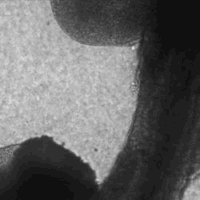
Crest cells migrating out of a gut
Neural Crest Cell Migration. Neural crest cells are an intrinsic part of who we are: they form our peripheral nervous system, the bones of our jaw, our skin pigments and our gut’s intrinsic innervation, the enteric nervous system (the « second brain »). Neural crest cell migration defects during embryogenesis give rise to a range of pathologies referred collectively as neurocristopathies. I work together with biologists and geneticists to understand the factors that control neural crest cells migration & differentiation.
Peristalsis. The intestine is a pump: it transports the food
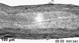
Propagating contractions in an embryonic gut
bolus by propagating electro-mechanical constrictions of the gut wall. This active transport mode is fundamental and common to most metazoans (pluricellular organisms): it has enabled them to grow by overcoming the size-limit imposed by simple diffusive food transport. Peristalsis starts very early on during embryogenesis: we are identifying the fundamental mechanisms underlying these early coordinated movements. This will allow us to better understand motility disorders.
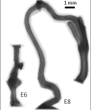
Gut evolution over 2 days of embryonic development
Growth. The adult intestine is on average 5m long , and only a few centimeters in diameter. These spectacular dimensions result from strong anisotropic growth during embryogenesis. I work on the physical and biochemical factors involved in gut growth. This research has both fundamental implications – understanding how organs grow – and applied medical perspectives for organ regeneration methods. As cancerous tumours often aberrantly resume embryonic organ growth programs in adults, we also hope our research can lead to a better understanding of tumor growth.
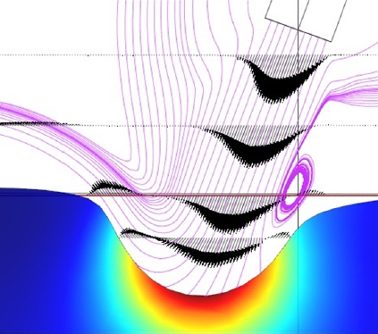 Methods. I am keen on developing new methods that fit our research needs. We have developed with V. Fleury a new elastography method for tissues and soft materials based on fluid-jet indentation, and introduced the use of the atomic force microscope to obtain elastography maps of embryonic tissue sections. Here is a non-exhaustive list fo the experimental methods we apply.
|
Microscopy |
Biology | Physics |
| Time-lapse Fluorescence Confocal Second Harmonic Generation Transmission & Scanning Electron |
Organ culture Immunohistochemistry Western blots PCR |
Micromechanical tool development
|
Before turning to embryonic development, I worked on crystal growth phenomena: Liesegang rings, colloidal crystals, heterogeneous nucleation and biomineralization. I keep a fond interest for these research areas. I am also head of the French International Young Physicist Tournament association, an experimental physics competition for high-school students. I have a keen interest in history of science and I am an amateur cellist.